Personalized tPCS Modeling and Neuronal Response Analysis
by IT’IS Foundation in Collaboration with AscenZionAnatomical and Electromagnetic Modeling
A population of high-resolution anatomical head models was used to capture inter-subject variability. The models account for conductivity contrasts between scalp and skull layers, dura, vasculature, gray and white matter, cerebrospinal fluid, and eye tissues. Sim4Life handles tissue property assignment according to the curated, traceable, and quality-assured IT'IS Tissue Properties Database .Electrodes were positioned according to the standardized 10–10 system for electroencephalography (EEG) recording (using artificial intelligence (AI)-based placement) and applied as boundary conditions for computing current-normalized electric potential and field distributions. Simulations were performed using Sim4Life's high-performance low-frequency electro-quasistatic solver, optimized for high-resolution medical device simulation in realistic anatomies – a requirement confirmed by a careful convergence study. A new Sim4Life Sample-Surface tool for generating Anchors, i.e., local coordinate systems, was used to sample exposure conditions throughout the cortex at neuroanatomically realistic depths corresponding to different neural populations.
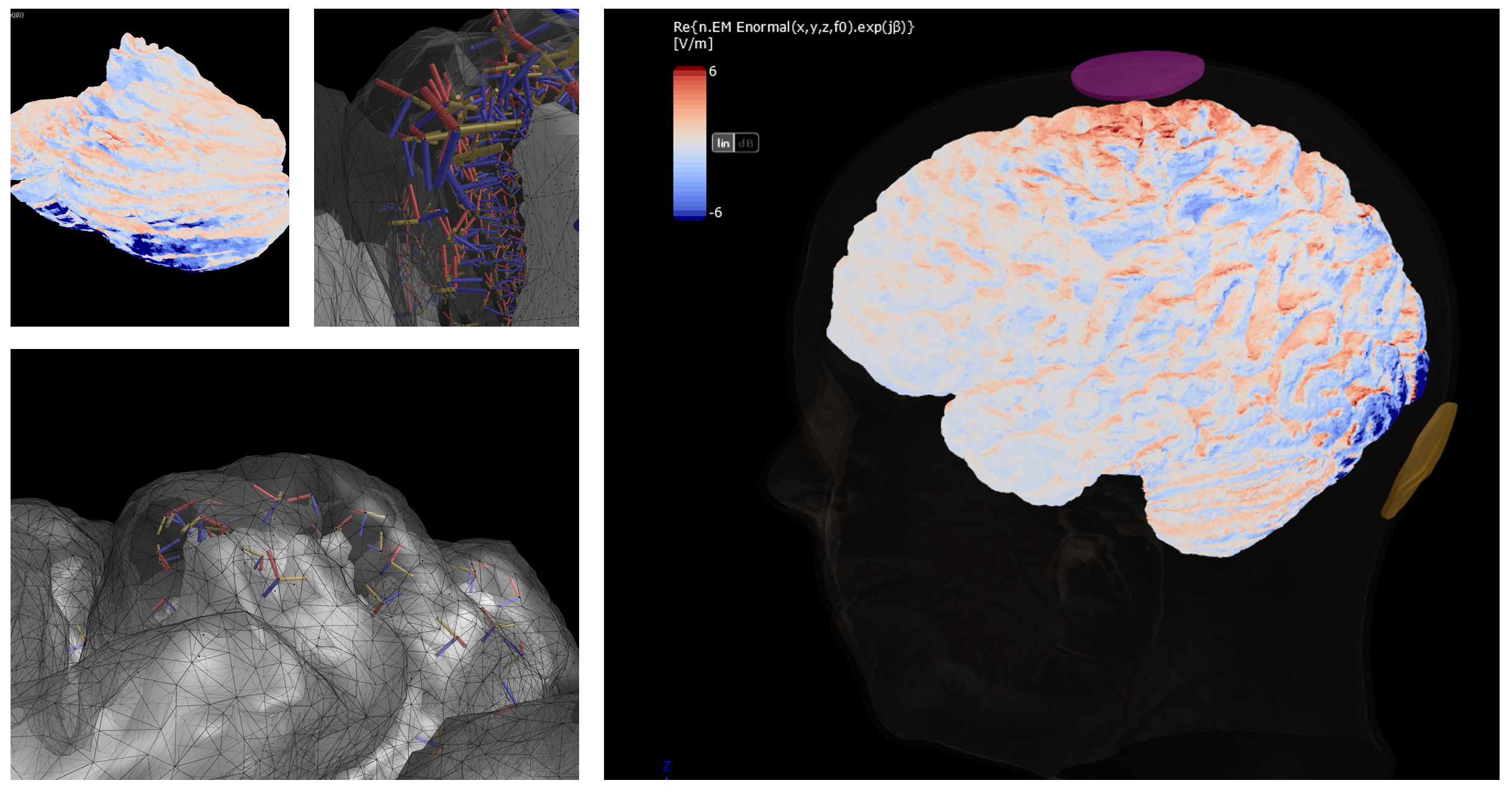
Electric field induced in the brain by electro-stimulation at 400Hz and visualized at the surface of the grey matter of the cerebrum and cerebellum. A subset of the sampled neurons, with their local coordinate systems, are shown inside the grey matter.
Neuron-Level Response Modeling
Morphologically and electrophysiologically detailed models of relevant cortical neurons, i.e., cerebellar Purkinje cells, Layer 5 pyramidal neurons and parvalbumin cells, were imported into Sim4Life from the well-established ModelDB repository. Sim4Life's T-Neuro module was then used to determine exposure-dependent subthreshold soma polarization, firing rate modulation, and suprathreshold stimulation limits.These response functions are pulse-shape-specific and were determined for three stimulation paradigms: classic alternating current stimulation, pulsed current stimulation with ideal pulse shapes, and pulsed current stimulation accounting for electrode-tissue-interface (ETI) distortion. For the latter, Sim4Life provides predefined equivalent circuit models for both thick and thin skin ETI.
These response functions were then combined with local exposure conditions at anatomically relevant cortical locations to produce cerebral and cerebellar maps of initial neural responses for each head anatomy.
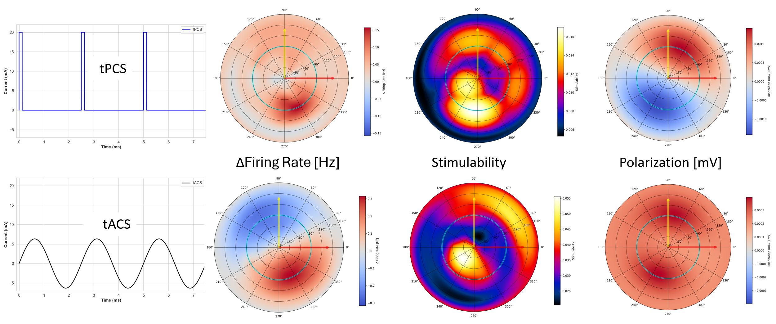
Orientation-dependent neural response functions used for “neuro-functionalization” of the head models, compared between tPCS (top row) and tACS (bottom row) at 400Hz. The polar maps summarize how each cell type’s response varies with electric field direction (elevation θ and azimuth φ) at a fixed peak field magnitude (same peak for tPCS and tACS). Columns (left→right) correspond to Purkinje cells, L5 pyramidal cells and PV interneurons. Shown metrics are ΔFiring Rate (Purkinje), Stimulability (inverse spiking threshold; higher means easier to excite), and Soma Polarization (peak “max” change in somatic membrane potential relative to no stimulation).
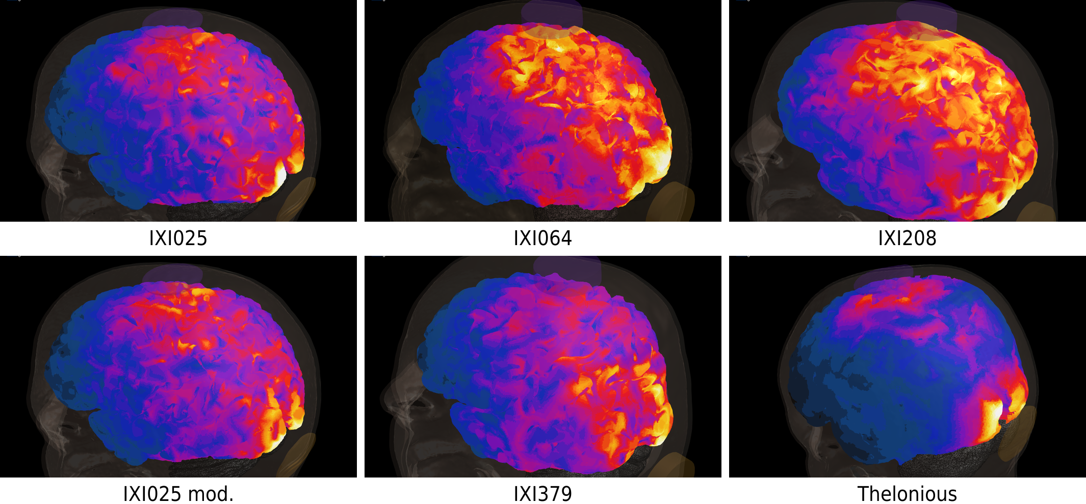
Cortical maps showing the spatial distribution of L5 pyramidal cell stimulability across different head models (IXI subjects and Thelonious) for tPCS. Colors indicate relative excitability, highlighting pronounced subject-specific differences in magnitude and spatial pattern driven by anatomical variability.
Inter-Subject Variability
Sim4Life's AI-powered segmentation tools enable the fully automatic generation of detailed, personalized head models (comprising 40 distinct tissues) directly from magnetic resonance imaging data, along with co-registration of a detailed brain atlas to identify structures relevant to brain stimulation. This made it practical to apply the complete modeling pipeline – from electrode configuration to neuron-level response estimation – across a population of anatomical head models. The resulting analysis quantified inter-subject variability in cortical exposures and neural responses, confirming the need for anatomically personalized stimulation planning.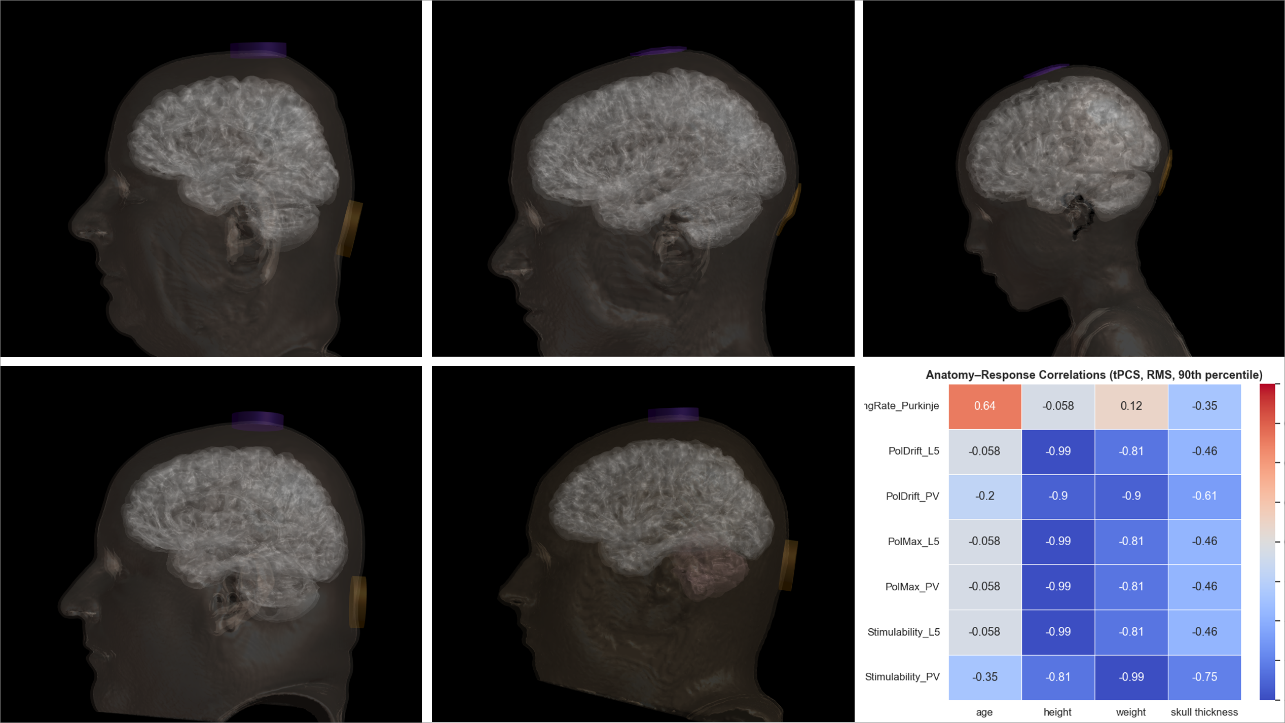
Spearman correlations (ρ) between anatomical properties and the 90th percentile of simulated QOIs under tPCS stimulation. Colors indicate direction and strength of association (centered at zero).